


Sunspots vs. Skin Cancer: How to Tell the Difference
If you have spent years enjoying summers at the beach or simply living without sunscreen, you likely have a few sunspots scattered across your face, chest, or hands. These flat, brown patches of hyperpigmentation are among the most common signs of cumulative sun exposure and, for the vast majority of people, are completely harmless. But because sunspots can sometimes resemble more serious lesions, it is natural to wonder where the line lies between a normal sunspot and an early sign of skin cancer. Understanding the difference is one of the most important things you can do for your long-term health, and it starts with a consultation with Dr. Michele Green.
Skin cancer is the most common cancer in the United States, and most cases are directly linked to ultraviolet light exposure from the sun or tanning beds. While sunspots themselves are not cancerous, they are markers of sun damage. The same UV rays that cause them can also trigger changes in melanocytes that lead to melanoma, basal cell carcinoma, or squamous cell carcinoma. These three cancers behave very differently: basal cell and squamous cell carcinomas tend to grow slowly and remain localized, while melanoma can spread quickly to other parts of the body if left untreated, making early detection especially critical. Warning signs to watch for include a mole that changes in size, shape, or color; a spot with irregular or blurred borders; a lesion that itches, bleeds, or fails to heal; or any new growth that looks different from the patient’s other moles and spots. These signs are often summarized by dermatologists like Dr. Green as the ABCDE rule (asymmetry, border irregularity, color variation, diameter, and evolution). Learning to recognize the subtle differences between a benign spot and a worrisome lesion can help catch a problem early, when it is most treatable. This is exactly the kind of guidance patients receive during a skin cancer screening with Dr. Michele Green, who combines visual inspection with dermoscopy and, when needed, biopsy to accurately distinguish sun damage from early malignancy.
Dr. Michele Green is a board-certified dermatologist based on the Upper East Side of Manhattan, with over 30 years of experience helping patients protect and rejuvenate their skin. Renowned for her expertise in medical and cosmetic dermatology, Dr. Green has been recognized by Castle Connolly, The New York Times, Super Doctors, and New York Magazine as one of New York City’s top dermatologists. Her patients trust her not only for sophisticated, natural-looking cosmetic results but also for her careful, experienced eye for identifying and managing skin cancer and other dermatologic conditions. Beyond treatment, she takes the time to help patients understand their skin, empowering them to recognize changes early and make informed decisions about their care.
How can you tell a sunspot from skin cancer?
At a glance, sunspots and certain skin cancers can look surprisingly similar, so self-checks alone are not always enough. A sunspot stays flat, uniformly tan or brown, and unchanged for months or years, while skin cancer lesions tend to evolve, changing color, growing, or becoming raised and tender. Melanoma can even develop within an existing mole, which is why comparing a spot to an older photo is often more telling than judging it on its own. A dermatologist can often distinguish the two with a handheld dermatoscope, but any spot in doubt should be evaluated. A biopsy provides a definitive answer. Dr. Michele Green combines visual inspection with dermoscopy to give patients clarity rather than leaving them to monitor an uncertain spot on their own.
One may ask, specifically, what melanoma looks like compared to a sunspot. Melanoma is the most dangerous form of skin cancer because it can spread quickly to other parts of the body if not caught early. A sunspot, by comparison, is flat, evenly pigmented, and remains consistent in color and shape over time. Melanoma, on the other hand, often appears as a dark, multicolored lesion with jagged or notched borders. It may show shades of black, brown, tan, red, or even white within the same spot, and it frequently grows or changes shape over weeks to months. Less commonly, melanoma can appear without pigment at all, presenting as a pink, red, or skin-colored bump, which makes it especially easy to overlook or mistake for a harmless irritation. Melanomas can also develop within an existing mole rather than appearing as a brand-new spot, which is another reason routine self-checks and professional skin exams are so valuable for early detection. When caught in its earliest stage, before it has spread beyond the top layer of skin, melanoma is highly treatable, which is why Dr. Green emphasizes that patients should never wait to have a changing or unusual spot evaluated.
Another common concern is whether sunspots itch, bleed, or hurt like skin cancer. A true sunspot is painless and remains flat and smooth, without itching, bleeding, or tenderness. Skin cancers, by contrast, often cause these symptoms, sometimes before any visible change appears. A lesion that bleeds without injury, crusts and reopens, itches persistently, or develops new tenderness or burning should be evaluated promptly. These symptoms can develop gradually, so patients sometimes mistake them for irritation from clothing or skincare products rather than a reason to see a dermatologist. Any persistent or unexplained symptom, however minor, is worth having checked by a board-certified dermatologist such as Dr. Michele Green.
Can a sunspot turn into skin cancer?
A genuine sunspot, composed of simple pigment buildup, does not itself transform into skin cancer. However, sunspots remain meaningful because they are visible evidence of significant cumulative sun exposure and damage, and patients with many sunspots are statistically at higher risk of developing skin cancer elsewhere on their bodies. In this sense, sunspots can serve as a warning sign, not because they become cancerous themselves, but because they reflect the same UV exposure that drives the development of melanoma, basal cell carcinoma, and squamous cell carcinoma. Dermatologists evaluating a patient with numerous sunspots will often examine the entire body, not just the pigmented areas, since the same sun exposure that created the visible spots may also be quietly contributing to changes elsewhere. Patients with extensive sun damage are encouraged to schedule more frequent skin checks with a board-certified dermatologist, such as Dr. Michele Green, who can establish a personalized screening schedule based on skin type, sun history, and overall risk factors. Since existing sunspots are a sign of past damage, daily broad-spectrum sunscreen with an SPF of 30 or higher is essential to prevent new spots from forming and to reduce the ongoing risk of skin cancer, making it one of the simplest and most effective tools a patient has for protecting their skin long term.
How fast does skin cancer typically grow compared to a sunspot?
A sunspot, once formed, is remarkably stable. It may fade slightly with treatment or darken further with additional sun exposure, but it generally does not change shape, develop new colors, or grow significantly over a short period. Skin cancers behave very differently. Melanoma can grow and spread within weeks to months, often showing visible changes in size, shape, color, or texture over a relatively short window. A mole or spot that appears asymmetrical, has an irregular or blurred border, contains multiple colors, or measures larger than a pencil eraser should be evaluated promptly, since these features are commonly associated with melanoma. Basal cell carcinoma and squamous cell carcinoma tend to progress more slowly, often over months to years. Yet, they still show ongoing growth rather than the static appearance of a benign lesion. These cancers may present as a persistent bump, a sore that does not heal, or a patch of skin that becomes scaly, tender, or prone to bleeding. Because early detection significantly improves treatment outcomes for all forms of skin cancer, any spot that is visibly larger than it was a few months ago, or that has changed in color, texture, or shape, warrants prompt attention. Regular skin checks, both self-exams and professional evaluations with a board-certified dermatologist like Dr. Green, remain the most reliable way to catch these changes early and distinguish a harmless sunspot from something that requires further evaluation.
What is the ABCDE rule for spotting skin cancer?
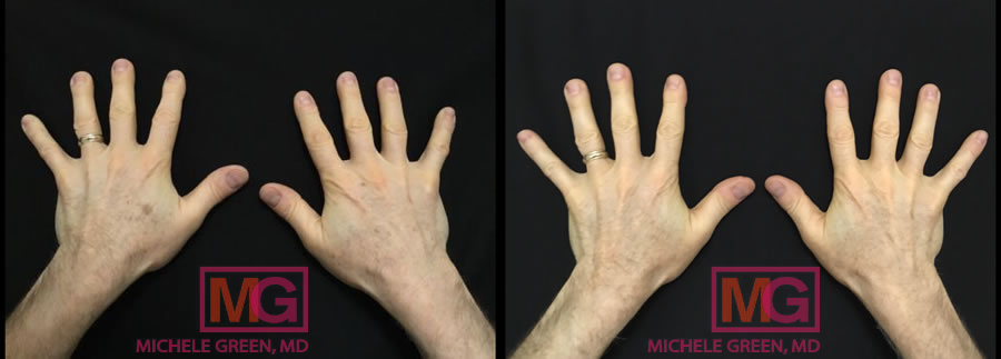
The ABCDE rule is a simple, memorable framework dermatologists use to help patients identify which spots warrant a closer look. The A stands for asymmetry, meaning one half of the spot does not match the other. This is a hallmark of irregular cell growth rather than the balanced, symmetrical shape seen in most benign spots. The B stands for border, since cancerous lesions often have ragged, blurred, or irregular edges rather than the smooth border typical of a sunspot. The C stands for color, as multiple shades of brown, black, red, or blue within a single lesion can be a red flag, since benign spots are usually a single, consistent shade throughout. The D stands for diameter, since spots larger than a pencil eraser (roughly 6mm) deserve extra attention. However, melanomas can sometimes be smaller, so size alone should never be the deciding factor in whether to seek an evaluation. The E stands for evolving, which captures any change in size, shape, color, or texture over time. Many dermatologists consider this the single most important warning sign, since even a spot that looks unremarkable on its own can be concerning if it is actively changing. Patients are often encouraged to perform a monthly self-check in good lighting, ideally with the help of a partner or a mirror for hard-to-see areas like the back and scalp, and to photograph any spots of concern so that subtle changes over time are easier to track. If a spot checks any of these boxes, it is time to schedule a skin cancer screening with a board-certified dermatologist, such as Dr. Michele Green.
When should you see a dermatologist about a spot?
Patients should schedule an appointment with a dermatologist whenever a spot is new, changing, or simply does not look or feel like the sunspots and moles they are used to. This includes any asymmetrical lesion with an irregular border, multiple colors, a diameter larger than a pencil eraser (6mm), or a spot that itches, bleeds, crusts over, or fails to heal, in keeping with the ABCDE rule of asymmetry, border irregularity, color variation, diameter, and evolution. These signs can be subtle at first, so a spot should not be dismissed simply because it seems small or unremarkable. Patients with fair skin, a history of sunburns, frequent tanning bed use, or a family history of skin cancer are at higher risk and should be especially vigilant. Annual full-body screenings are strongly recommended, even without a specific worrisome spot. Patients who are concerned or unsure about a particular spot are encouraged to consult with Dr. Green, who can help determine whether a lesion is simply a harmless sunspot or something that warrants closer evaluation.
Most dermatologists, including Dr. Green, recommend an annual full-body skin exam for patients with significant sun damage or numerous sunspots. Those at higher risk may benefit from visits every 6 months. During these exams, Dr. Green checks the skin head to toe, including often-overlooked areas such as the scalp, ears, back, and between the toes, because skin cancer can develop in both sun-exposed and non-sun-exposed areas. She may also use dermoscopy to get a closer look at any atypical spots. Patients are encouraged to report any spots that have changed or developed new symptoms, such as itching or bleeding, since regular exams help catch skin cancer early and make it easier to track changes over time.
Frequently Asked Questions (FAQs) about Sunspots vs Skin Cancer
What’s the difference between a sunspot and a mole?
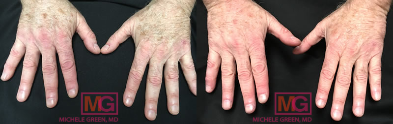
Sunspots are among the most common cosmetic concerns dermatologists see, but they are often mistaken for far more serious conditions, from moles to melanoma. Understanding what separates a harmless spot from one that needs medical attention can make the difference between early treatment and a missed diagnosis.
A sunspot, also called an age spot or liver spot, develops when years of sun exposure prompt melanocytes to produce excess pigment in a concentrated area, resulting in a flat, evenly colored brown or tan patch. These spots tend to appear on areas that receive the greatest cumulative sun exposure over a lifetime, including the face, hands, shoulders, and chest, and they become more numerous with age. A mole, by contrast, is a cluster of melanocytes that can be present from birth or develop over time. Most new moles form during childhood and adolescence, with fewer appearing after age forty. Moles tend to be more uniformly round and can be flat or slightly raised. While they are not necessarily related to sun exposure, moles on sun-exposed skin can still be influenced by UV damage. Both are usually benign, but moles warrant closer attention because they are more likely than sunspots to develop into melanoma, particularly when a mole looks atypical, runs in the family, or shows any of the changes described by the ABCDE warning signs. Patients with a higher number of moles or a personal or family history of melanoma are often advised to schedule skin evaluations more frequently.
What’s the difference between actinic keratosis and a sunspot?
Actinic keratosis is another spot often mistaken for sun damage, but unlike a sunspot, it is a precancerous lesion that requires medical attention. A sunspot is flat and smooth, driven purely by hyperpigmentation, while actinic keratosis typically appears as a rough, scaly, or crusty patch that can feel like sandpaper. This texture is often easier to detect by touch than by sight in the earliest stages. These patches range from skin-toned to reddish-brown and commonly appear on areas with the highest lifetime sun exposure, such as the face, ears, scalp, forearms, and backs of the hands. Left untreated, a significant proportion of these lesions eventually progress to squamous cell carcinoma, which is why dermatologists rarely recommend simply watching and waiting once one is identified. Because actinic keratosis and sunspots can look similar in their early stages, any new rough or scaly spot deserves evaluation rather than being assumed. Dr. Michele Green offers a range of treatment options once actinic keratosis is identified, including topical medications such as Efudex and Photodynamic Therapy (PDT). She tailors the approach to the severity of each patient’s lesions.
What does basal cell carcinoma look like vs. a sunspot?
Basal cell carcinoma is the most common skin cancer, affecting approximately two million people each year. It arises from the basal cells, which regenerate new skin as older cells die off. In most cases, it develops on areas of the skin with the greatest cumulative sun exposure, such as the face, neck, ears, back, and scalp, though it can occur anywhere on the body. Rather than a flat, brown patch, basal cell carcinoma often appears as a pearly or waxy bump, a flat, flesh-colored or brown, scar-like lesion, or a sore that bleeds, crusts over, and never seems to heal fully. Small blood vessels may be visible on the surface, and the center can appear slightly indented or shiny, details that are easy to miss without a close, professional look. It tends to grow slowly and rarely spreads to other organs, which can lead patients to underestimate it. Left untreated, it can still invade deeper into surrounding tissue and cause significant local damage. This is why early detection and treatment are so important. When caught early, basal cell carcinoma is highly treatable. When Dr. Green diagnoses basal cell carcinoma, treatment typically involves excision, Mohs surgery, or electrodesiccation, depending on the lesion’s size and location. All three approaches have excellent success rates, especially when detected early through frequent skin checks with a board-certified dermatologist like Dr. Green.
Is squamous cell carcinoma ever mistaken for a sunspot?
Squamous cell carcinoma is the second most common form of skin cancer, arising in the squamous cells that make up the outer layers of the epidermis. It’s driven primarily by cumulative UV exposure, though it can also arise from chronic wounds, scars, or long-standing inflammation. In its earliest stages, squamous cell carcinoma can be mistaken for a sunspot or a stubborn patch of sun damage. Over time, however, it typically develops into a firm, red, scaly bump or a flat, crusted lesion and may feel tender or itchy, unlike a sunspot. The surface can also become wart-like or ulcerated, and some lesions grow steadily larger over weeks or months rather than remaining static. Squamous cell carcinoma often arises from untreated actinic keratosis, underscoring the importance of never ignoring precancerous spots. Unlike basal cell carcinoma, squamous cell carcinoma carries a higher, though still relatively low, risk of spreading to nearby lymph nodes or other tissues if left untreated for an extended period, making timely evaluation especially important. If found early, squamous cell carcinoma is highly treatable, often with in-office excision, electrodesiccation, or Mohs surgery, though more advanced cases may require radiation therapy.
Are raised or bumpy sunspots more likely to be cancerous?
A true sunspot is typically flat, with a smooth surface that blends into the surrounding skin, aside from its pigmentation. A spot that has become raised, bumpy, or developed a different texture from the surrounding skin is more suspicious and should be examined by a board-certified dermatologist like Dr. Green, as it may be an early sign of basal cell carcinoma, squamous cell carcinoma, or actinic keratosis. A rough, scaly, or crusty texture is particularly notable, as actinic keratoses often feel like sandpaper to the touch even before they are visually obvious, meaning a change may be detected by feel before it can be seen. Basal cell and squamous cell carcinomas may appear as a pearly bump, a firm nodule, a scaly patch, or a sore that does not heal, and they can sometimes be mistaken for a pimple, insect bite, or scar that is simply taking a long time to resolve. Because these changes in texture can be subtle at first and easy to mistake for dry skin, patients who notice any new roughness, thickening, or elevation within an existing sunspot should have it evaluated by Dr. Green rather than waiting to see if it resolves on its own, since early evaluation allows for simpler and more effective treatment.
Do sunspots have irregular borders like melanoma can?
No, sunspots almost always have smooth, well-defined borders that clearly separate the lesion from the surrounding skin. An irregular, blurred, or notched border is a hallmark of melanoma and part of the ABCDE rule (Asymmetry, Border irregularity, Color variation, Diameter greater than 6mm, and Evolving appearance) that dermatologists use during a skin exam. Sunspots also tend to be uniform in color, typically appearing as a single shade of tan, brown, or black throughout the entire lesion. In contrast, melanoma often displays several colors within the same lesion, including shades of brown, black, red, white, or blue, sometimes in an uneven or patchy distribution. This color variation can be one of the earliest visual clues that a spot is not a typical sunspot, even before other features become apparent. Any spot that fails to meet these criteria, even if it otherwise resembles a sunspot in other respects, should be evaluated promptly by Dr. Green. Patients who are unsure how to assess their own spots against the ABCDE rule are encouraged to bring their questions to a skin check rather than attempt to self-diagnose, since subtle warning signs can be difficult to distinguish without professional training and dermoscopy. This technique allows for magnified, detailed examination of pigmented lesions.
Can sunspots be a sign you’re at higher risk for skin cancer?
While sunspots are not cancerous, having many of them indicates a history of significant UV exposure, the leading risk factor for skin cancer. Patients with extensive sunspots should be especially diligent about sunscreen use and routine skin checks. Because sunspots and early skin cancers can look similar, a higher sunspot count signals to Dr. Green that a patient’s skin has accumulated years of sun damage, warranting closer monitoring. These patients are encouraged to perform regular self-exams at home, looking for any new spots or changes in size, shape, color, or texture of existing ones. Daily use of a broad-spectrum sunscreen with an SPF of 30 or higher, reapplied every two hours during sun exposure, remains one of the most effective ways to prevent additional sun damage and reduce future skin cancer risk. Dr. Green’s own MGSKINLABs Hydrating SPF 50 sunscreen was formulated with this level of daily protection in mind, offering patients a broad-spectrum option that fits easily into a daily skincare routine. Annual or biannual skin checks with Dr. Green allow for professional evaluation of any concerning areas, along with dermoscopic examination when needed, so that any changes can be caught and addressed as early as possible.
What’s the difference in survival rates if a “sunspot” turns out to be melanoma?
Early detection makes an enormous difference. Melanoma detected at its earliest stage has a very high survival rate, often exceeding 99 percent when caught before it spreads beyond the skin’s surface. By contrast, melanoma that is allowed to grow and spread becomes significantly more difficult to treat and carries a far more guarded prognosis. This stark difference in outcomes is largely a matter of timing rather than the aggressiveness of any individual case, which is why vigilance matters so much. This is precisely why any spot that resembles a sunspot but shows worrisome changes should never be dismissed without evaluation, even if it seems minor or the change has been gradual. Waiting to see if a spot resolves on its own can allow a treatable condition to progress unnecessarily. Patients who notice a suspicious spot or are unsure whether a mark is a harmless sunspot or something more concerning can consult with Dr. Green to better understand what they’re seeing and determine the appropriate next steps, whether that means simple reassurance, closer monitoring, or a biopsy for a definitive diagnosis.
Can you get a biopsy for a sunspot just to be safe?
If a sunspot has unusual features or you simply want peace of mind, Dr. Green can perform a quick, in-office biopsy. A small sample of the lesion is removed and sent to a lab, providing a definitive diagnosis and ruling out skin cancer. The procedure is minimally invasive and typically takes just a few minutes. It is performed under local anesthesia to keep patients comfortable throughout, and most people report only mild pressure rather than pain. Depending on the size and location of the lesion, Dr. Green may use a shave biopsy, which removes the top layers of skin, or a punch biopsy, which takes a small, deeper cylindrical sample. The choice of technique depends on how deep the suspicious tissue appears to be and where on the body the spot is located. Most patients experience little to no downtime afterward, aside from minor care of the biopsy site as it heals, such as keeping the area clean and covered for a few days. Results are usually available within a week, at which point Dr. Green will discuss the findings and any recommended next steps. Because a biopsy removes any guesswork, it remains the gold standard for distinguishing between a harmless sunspot and a more serious skin condition, offering patients clarity and reassurance that visual examination alone cannot provide.
How to evaluate your sun spots today?
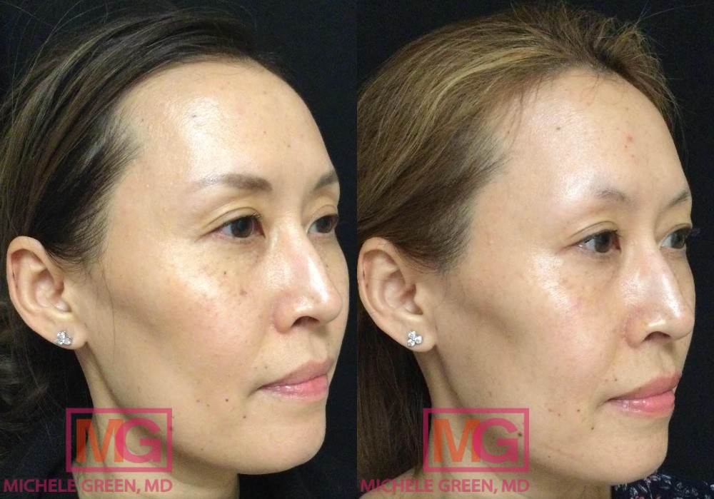
Sunspots are a common, generally harmless byproduct of sun exposure, and the vast majority of patients who develop them will never need anything more than reassurance or optional cosmetic treatment. Still, they share enough visual similarities with certain skin cancers that self-diagnosis is never a safe substitute for a professional opinion. Even experienced patients who have lived with sunspots for years can find it difficult to tell when a familiar spot has crossed the line into needing attention. By understanding the ABCDE rule, watching for symptoms such as itching, bleeding, or a change in texture, and recognizing the differences among sunspots, moles, actinic keratoses, and true skin cancer lesions, you can be a more informed advocate for your skin health. This knowledge does not replace professional evaluation, but it does make it easier to know when a spot deserves a closer look. When in doubt, the safest course of action is early detection through a thorough skin exam with a qualified dermatologist, such as Dr. Michele Green, whose experience helps patients move forward with clarity rather than uncertainty.
Dr. Michele Green is a world-renowned, board-certified dermatologist based on the Upper East Side of Manhattan, New York City, with over 25 years of experience providing the most advanced and effective cosmetic and medical treatments. She helps patients protect their skin and treats everything from routine sun damage and hyperpigmentation to complex skin cancer cases, often in coordination with Mohs surgeons when surgical intervention is needed. She is consistently recognized by Castle Connolly, Super Doctors, and New York Magazine as one of the best dermatologists in New York City and has helped countless patients. Her thorough, attentive approach to skin exams has made her a trusted resource for patients seeking clarity and reassurance about their sunspots, moles, and other lesions. To schedule a comprehensive skin cancer screening or consultation, call Dr. Michele Green’s Upper East Side office today at 212-535-3088 or contact us online.


