
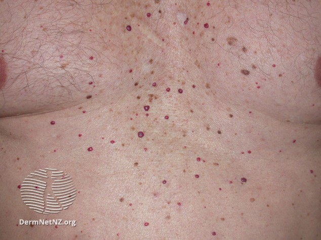
Cherry Angioma (Cherry Hemangioma)
A cherry angioma is a common, benign vascular lesion made up of proliferating endothelial cells, the cells that line the inside of blood vessels. Cherry angiomas are typically dome-shaped red papules or macules. Factors like aging, pregnancy, genetics, and chemical exposure can contribute to the development of cherry angiomas. Since the exact cause of cherry angiomas remains unknown, there isn’t a method of prevention that is 100% guaranteed to work. Although cherry angiomas can not fully be prevented, they can easily be removed by a board-certified dermatologist like Dr. Green without downtime or discomfort to reveal a clear complexion.
Cherry angiomas do not go away on their own and are usually removed for cosmetic reasons. Effective non-invasive treatment options for cherry angiomas include the V-Beam laser, cryosurgery, shave excision, IPL, and electrodesiccation. Cherry angiomas are usually benign and of little concern; however, if a lesion changes in appearance (size, color, or shape) or starts to bleed, it may be an early indication of skin cancer. Any dermal lesions that are evolving or associated with any kind of irritation should be evaluated by an experienced board-certified dermatologist, such as Dr. Michele Green in NYC. If necessary, Dr. Green can biopsy skin lesions for laboratory assessment to rule out skin cancer.
Dr. Michele Green is an internationally renowned board-certified dermatologist with over two and a half decades of experience providing some of the world’s most discerning individuals with the best non-invasive cosmetic treatments, including cherry angioma removal. She customizes each patient’s treatment plan to suit their particular concerns and aesthetic goals best. Dr. Green is consistently identified as one of New York’s best dermatologists by Castle Connolly, Super Doctors, New York Magazine, and the New York Times for her dedication to her patients and expertise.
What is a cherry angioma?
A cherry angioma, also known as a Campbell de Morgan spot or senile angioma, is a common skin growth containing an abnormal proliferation of tiny lymphatic or blood vessels. These growths are benign and do not cause any discomfort. Cherry angiomas are extremely common and found with increasing age, as around 50% of adults over 30 years old and 75% of adults over 75 years old have cherry angiomas. There is no known cause for why cherry angiomas appear, although age and genetics may play a factor. Cherry angiomas are not a sign of cancer or other medical conditions. However, they can look similar to other skin lesions, and the differential diagnosis for cherry angiomas includes skin cancers such as nodular basal cell carcinoma and amelanotic melanoma; if you notice any changes in the characteristics of a cherry angioma or are unsure if you have a cherry angioma, consult a board-certified dermatologist such as Dr. Green for proper assessment and diagnosis.
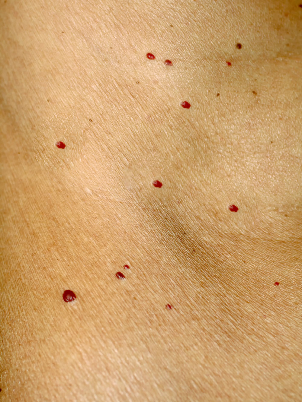
What does a cherry angioma look like?
Cherry angiomas, as the name suggests, are cherry-red in appearance due to the collection of small lymphatic and blood vessels. Cherry angiomas, however, can also have a bluish or purplish hue. Some cherry angiomas are flat, while others are dome-shaped and slightly raised above the skin. They vary in size from about 2 to 4 mm and can be circular or oval-shaped.
Cherry angiomas are unique in appearance but can be confused with spider angiomas or angiokeratomas. With spider angiomas, their color is not as bright red and fades when touched. Angiokeratomas are typically harder to the touch and have a rough or warty appearance. On the other hand, cherry angiomas are typically bright red in appearance, are softer to the touch, and may occasionally bleed and become irritated, especially if aggravated by clothing or other external factors. An experienced healthcare professional, such as board-certified dermatologist Dr. Green, however, has the expertise required to tell the difference between these lesions.
What is the cause of cherry angiomas?
While the exact cause of cherry angiomas is unknown, research and anecdotal evidence suggest they may develop due to age, hormonal changes, and genetics. Cherry angiomas are a prevalent condition in adults over 30 years of age, with incidence increasing in aging, as over 75% of individuals over 75 years old have cherry angiomas. These lesions additionally seem to get larger and more pronounced with age. There is also a high incidence of cherry angiomas in pregnant women, likely due to fluctuations in hormone levels during pregnancy. Genetics is another factor believed to cause cherry angiomas, as many individuals with cherry angiomas have family members similarly affected. In some cases, multiple cherry angiomas may develop at once due to health conditions such as herpesvirus-* (HHV8) or immunosuppressant usage – these cases are referred to as eruptive cherry angiomas. Other factors, such as liver dysfunction, elevated levels of bromide within the body, and excessive sun exposure, may also play some role in cherry angioma development.
What is the difference between cherry angiomas and cherry hemangiomas?
Cherry angiomas and cherry hemangiomas are common, benign skin growths that are very similar in appearance but are made of different cells. While cherry angiomas are made of lymphatic or blood vessels, cherry hemangiomas are made of only blood vessels. Cherry hemangiomas may appear in infancy or childhood, whereas cherry angiomas usually affect adults. The term ‘cherry’ simply refers to the color of these skin growths, which can appear as bright cherry-red to purple. Both types of skin lesions are usually between two and four millimeters and may be smooth and flat or raised and dome-shaped. Cherry angiomas and cherry hemangiomas do not go away on their own. While cherry angiomas and hemangiomas normally don’t require removal for medical purposes, many individuals choose to have them treated for cosmetic reasons. Dr. Michele Green offers a number of treatment options for removing cherry angiomas and hemangiomas without discomfort, downtime, or residual scarring. When you consult with Dr. Green, she will work with you to create a personalized treatment plan for achieving and maintaining a clear, even-toned complexion that lasts.
Can cherry angiomas be removed? How to remove a cherry angioma
Yes. Cherry angiomas are usually removed for cosmetic reasons, as cherry angiomas are not an indication of any underlying health issues or medical conditions. Many people who have cherry angiomas may choose to have them removed if they are unhappy with their cosmetic appearance. There are times, however, when the removal of a cherry angioma is needed due to irritation or constant bleeding. Effective treatment options for cherry angiomas include the V-Beam laser, cryosurgery, shave excision, IPL, and electrodesiccation.
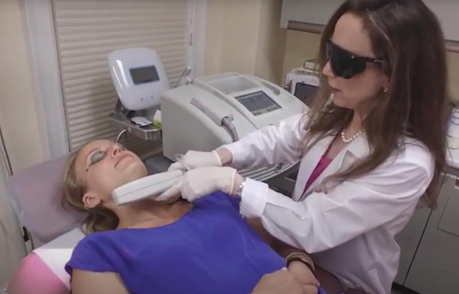
V-Beam pulsed dye laser treatment of cherry angiomas
The V-Beam laser, a pulsed dye laser, is one of the most effective treatments for removing cherry angiomas. The V-Beam is the gold standard when it comes to eliminating red pigment from the skin. The V-Beam operates on a specific wavelength of light that is absorbed by red pigment while surrounding skin cells are left untouched. Dr. Green uses this laser therapy to reduce visible blood vessels, stretch marks, bruising, active acne, post-inflammatory hyperpigmentation, symptoms of rosacea, and leg veins, among other skin conditions. The V-beam laser works by delivering quick pulses of laser light energy to destroy the cherry angioma, focusing the energy on the red capillaries inside the angioma. Before each laser pulse, a burst of cool air is emitted from the V-Beam’s Dynamic Cooling Device, which enhances patient comfort during the laser treatment. Patients may require several treatment sessions to obtain their final ideal cosmetic results.
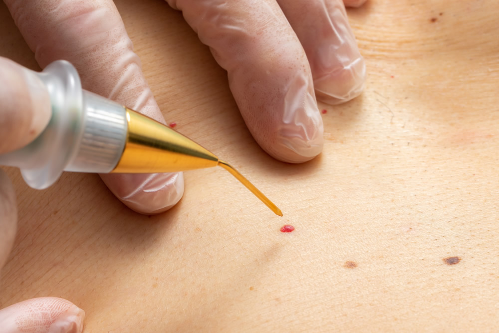
Cryosurgery for cherry angioma removal
Cryosurgery is another effective, non-invasive treatment option for removing cherry angiomas. This procedure was once very popular in dermatology. However, its popularity has diminished over the years in favor of other removal treatment options like the V-Beam or electrodesiccation, as it causes tissue damage and can take longer to heal from. Cryotherapy should only be performed by a certified dermatologist, such as Dr. Green in NYC. Cryotherapy entails the use of liquid nitrogen. Liquid nitrogen needs to be stored carefully as it can be combustible. To remove the cherry angiomas, the liquid nitrogen is gently applied onto a cotton swab; the swab is then gently pressed against the cherry angioma. The liquid nitrogen freezes the area on contact, which can sometimes cause a blister. The blister then heals by forming a scab. Once the scabs fall off, it should remove the cherry angioma, which would have dried up from the freezing.
Shave Excision for cherry angioma treatment
Shave Excision is an unpopular form of treatment for cherry angiomas due to the possibility of scarring. With an excision, the area is prepped, usually with a local anesthetic, and then a scalpel is used to biopsy the lesion. There can be downtime with the shave removal as the wound needs time to heal properly. This method may be used to confirm the diagnosis of a vascular lesion if it is uncertain.
Intense Pulsed Light (IPL) for cherry angiomas
Intense Pulsed Light (IPL) is an effective light treatment for the removal of cherry angiomas. Also known as the Photo Facial laser, the IPL works to reduce the redness and vascularity present in hemangiomas. The IPL emits light energy, which is absorbed deep into the dermis. Unlike most other lasers, IPL uses 560 wavelengths to target the treatment area. After the treatment, the collection of blood from the dilated vessels dissipates a few days after treatment, leaving clear skin. There may be a faint area of discoloration in the skin in the area where the hemangioma was previously located. This discoloration will fade over time with the use of proper skin care and sunscreen in sun-exposed areas.
Electrodesiccation of cherry angiomas
Electrodesiccation, also known as electrocautery, is another treatment often used to remove cherry angiomas, pre-cancerous lesions, skin tags, skin cancer, and other benign skin growths. Although this treatment is quick, some individuals may experience some slight discomfort during the procedure. Depending on your pain threshold, your dermatologist may recommend applying a topical anesthetic to the treatment area for 30-45 minutes before having the treatment performed. The topical anesthetic numbs the area before electrodesiccation so that you will not experience any discomfort during the treatment. The hyfrecator machine uses a tiny needle that heats up and destroys the blood vessels upon contact. Electrodesiccation treatment typically causes the skin to scab, which heals in a few days. It is important to refrain from prematurely picking the scab and allowing the skin to heal naturally, as forcing the scab can cause hypopigmentation or scarring. It is advised to keep the treated area covered with an anti-bacterial ointment as the scab heals on its own.
Can cherry angiomas go away?
Cherry angiomas do not go away on their own. A board-certified dermatologist, such as Dr. Michele Green in New York City, can remove cherry angiomas. There are several treatment options available at Dr. Green’s private dermatology office that can remove cherry angiomas without any downtime or discomfort. Cherry angiomas are usually removed for cosmetic reasons, as they are benign vascular lesions that do not require medical treatment. Once cherry angiomas have been treated, it can take 1-4 weeks to disappear, depending on the treatment option used for removal. When you have cherry angioma removal with Dr. Green at her private dermatology office in NYC, she will provide you with all of the pertinent aftercare information required to achieve the best results and let you know what to expect in terms of how long it will take for the lesions to go away after having treatment.
How to prevent cherry angiomas
Since the exact cause of cherry angiomas remains unknown, there isn’t a method of prevention that is 100% guaranteed to work. However, some general recommendations can help best prevent cherry angiomas. Limiting sun exposure and wearing proper sun protection is always recommended to prevent skin cancer and photodamage. It is also thought that limiting sun exposure will help protect against cellular changes that lead to the proliferation of endothelial cells that line blood vessels, which ultimately form cherry angiomas. Additionally, some treatments have been linked to the development of cherry angiomas. For example, one potential side effect of topical nitrogen mustard, a treatment for vitiligo, is the formation of cherry angiomas. Although cherry angiomas can not fully be prevented, they can easily be removed by a board-certified dermatologist like Dr. Green.
What causes red angiomas or red moles?
Angiomas are benign growths made up of proliferating endothelial cells, the cells that line the inside of blood vessels. The three most common types of angiomas are cherry angiomas, spider angiomas, and angiokeratomas. Spider angiomas are red or purple marks on the skin that resemble the body and legs of a spider and are caused by dilated or widened capillaries near the skin’s surface. Angiokeratomas are hard bumps under the skin that are red, blue, purple, or black due to an enlarging or rupturing of capillaries near the skin’s surface.
The exact cause of angioma formation is unknown, although research suggests genetics, hormones, and aging may play a factor. Many individuals with cherry angiomas have similarly affected family members and see increased development of cherry angiomas with age. Spider angiomas may be caused by increased levels of estrogen, as there is a high incidence of spider angiomas in individuals who are pregnant, going through puberty, and taking oral contraceptives. Spider angiomas may also be linked in some way to the liver, as multiple spider angiomas on the body may be a sign of an underlying liver disease. Angiokeratomas can be caused by vascular malformations, chronic irritation, or conditions that lead to increased pressure on blood vessels.
Are cherry angiomas dangerous?
No! Cherry angiomas are usually benign, harmless, and of little concern. These cutaneous vascular proliferations typically do not require any treatment; however, if you develop new lesions or red spots or notice a change in the appearance of a red mole, you should consult a dermatologist to determine if the growth is, in fact, a cherry angioma or another type of lesion or skin cancer. An expert dermatologist such as Dr. Michele Green will be able to confirm if the new growth or clinical changes in appearance warrant further evaluation and treatment. If necessary, a biopsy of the lesion may be performed and sent to a laboratory for histological review to rule out skin cancer or other dermatological conditions. If you are concerned about a new or changing skin lesion, it is always best to contact a board-certified dermatologist like Dr. Michele Green to get the best medical advice.
Can Cherry Angiomas Itch?
Cherry angiomas do not itch. These benign vascular lesions are asymptomatic. If your skin feels itchy near cherry angiomas, it may be due to conditions like dry skin, psoriasis, acne, or an allergy. Be careful not to scratch cherry angiomas, as doing so could result in bleeding, discomfort, infection, and scarring. If you are experiencing skin irritation or have noticed a lesion that bleeds or has changed in size, shape, or color, schedule a consultation with Dr. Green, who can assess it. Depending on her evaluation, a biopsy may be performed for histological review to rule out skin cancer and other conditions.
Do Cherry Angiomas Hurt?
No. Cherry angiomas are a benign condition that usually goes unnoticed. There are times, however, when cherry angiomas may cause some discomfort. If Cherry angiomas become irritated from excessive rubbing, rupture, or bleeding, they can be annoying and uncomfortable.
Are cherry angiomas cancerous?
No. Cherry angiomas are not cancerous. However, if cherry angiomas change in appearance (size, color, or shape) or start to bleed, it may be an early indication of skin cancer. Any dermal lesions that are evolving or associated with any kind of irritation should be evaluated by an experienced board-certified dermatologist, such as Dr. Michele Green in NYC. If necessary, Dr. Green can biopsy skin lesions for laboratory assessment to rule out skin cancer.
How to stop cherry angiomas from bleeding
Cherry angiomas often present as raised, dome-shaped red papules or macules with a smooth, flat surface. These vascular lesions can easily be scratched, potentially resulting in bleeding. To stop a cherry angioma from bleeding, clean the area, apply antibacterial ointment to the lesion, and cover it with a bandage. While cherry angiomas are usually nothing to worry about, schedule an appointment with a board-certified dermatologist if a skin lesion starts to bleed. Dr. Green is an internationally renowned board-certified dermatologist with over two and a half decades of experience treating some of the world’s most discerning men and women from her private, boutique dermatology office in Manhattan’s Upper East Side neighborhood. She is consistently identified as one of NYC’s best dermatologists by Super Doctors, Castle Connolly, and New York Magazine for her dedication to her patients and expertise.
Can you pop a cherry angioma?
No! Do not attempt to pop a cherry angioma. Trying to remove a cherry angioma on your own could result in bleeding, pain, infection, and scarring. Instead, schedule an appointment with a board-certified dermatologist like Dr. Green in New York City. Dr. Green can safely remove cherry angiomas without discomfort or downtime to reveal a clear complexion. The treatment options offered at Dr. Green’s office for cherry angioma removal are non-invasive and do not result in scarring, ensuring that you get the best results.
Why am I getting cherry angiomas?
Cherry angiomas are common benign vascular tumors that can affect adolescents and adults, although they tend to increase in size and number with aging. Most people affected by cherry angiomas are over 30 years old. The exact cause remains unknown, but factors like aging, pregnancy, chemical exposure, and genetic mutations can contribute to the development of cherry angiomas. Exposure to treatments with topical nitrogen mustard and bromides has been linked to the formation of cherry angiomas. Sun exposure is thought to contribute to cellular changes that lead to the proliferation of endothelial cells and, subsequently, cherry angiomas.
Why do I have so many cherry angiomas?
Aging is a big contributing factor in getting cherry angiomas, as the size and number of cherry angiomas tend to increase dramatically after the age of 40. While it is considered normal to develop cherry angiomas over time gradually, the sudden development of many cutaneous skin lesions may be an indication of an internal issue. If you have suddenly developed many cherry angiomas in a short period, you should consult a physician.
How to get rid of cherry angiomas at home
Do not attempt to remove cherry angiomas at home. There is no scientific evidence indicating the efficacy of home remedies in the removal of cherry angiomas. Attempting to remove cherry angiomas on your own can result in bleeding, infection, and scarring. Dr. Green can utilize one of the non-invasive treatment options available at her private dermatology office to remove your cherry angiomas without downtime, discomfort, or a residual scar.
What is the differential diagnosis for a cherry angioma?
An experienced board-certified dermatologist like Dr. Michele Green in NYC can diagnose cherry angiomas with a visual exam. Cherry angiomas are benign vascular lesions, but not every vascular lesion on the body is a cherry angioma. The differential diagnosis for cherry angiomas includes amelanotic melanoma, nodular basal cell carcinoma (BCC), pyogenic granulomas, and angiokeratomas. A biopsy of the skin lesion can be performed if there is uncertainty about the diagnosis. A cherry angioma is composed of venules in a thickened papillary dermis.
How to get started with cherry angioma treatment today
A cherry angioma or cherry hemangioma is a common, benign vascular lesion made up of proliferating endothelial cells, the cells that line the inside of blood vessels. Since the exact cause of cherry angiomas remains unknown, there isn’t a method of prevention that is 100% guaranteed to work. Although cherry angiomas can not fully be prevented, they can easily be removed by a board-certified dermatologist like Dr. Green without downtime or discomfort. Cherry angiomas are usually removed for cosmetic reasons, and effective treatment options for cherry angiomas include the V-Beam laser, cryosurgery, shave excision, IPL, and electrodesiccation.
Dr. Michele Green is an internationally renowned board-certified dermatologist with over two and a half decades of experience providing some of the world’s most discerning individuals with the best non-invasive cosmetic treatments, including cherry angioma removal. She customizes each patient’s treatment plan to suit their particular concerns and aesthetic goals best. Dr. Green is consistently identified as one of New York’s best dermatologists by Castle Connolly, Super Doctors, New York Magazine, and the New York Times for her dedication to her patients and expertise. To schedule a consultation, contact us online today or call Dr. Michele Green’s NYC-based office at 212-535-3088